VITEYE BUNDLE: Dermascope smartphone attachment + App
VITEYE BUNDLE: Dermascope smartphone attachment + App
Couldn't load pickup availability
Share
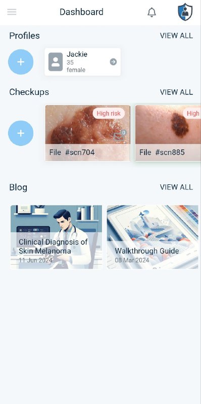
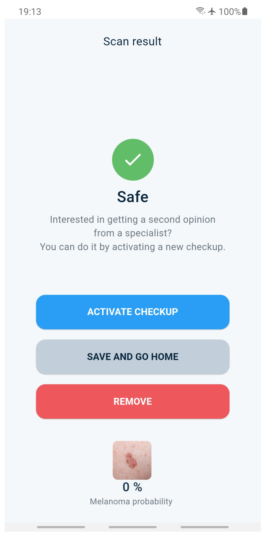
Viteye is a software application designed to aid in the early diagnosis of skin melanoma. It uses neural networks to analyze images of pigmented neoplasms and provide a probability of melanoma diagnosis.
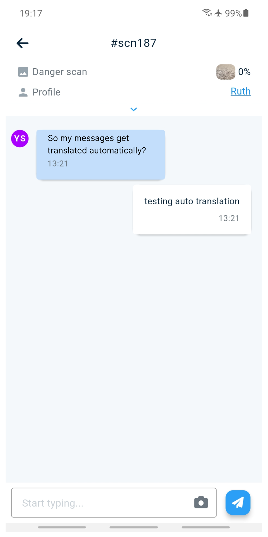
The application is intended for use by doctors, patients, and individuals seeking to enhance their expertise in melanoma diagnosis. It allows doctors and individuals to capture and analyze images of suspicious moles and to obtain opinions from other medical professionals.
The application is not intended to replace a doctor’s diagnosis, but rather to support the decision-making process and encourage early detection and treatment of skin melanoma.
Please visit our website to learn more about Viteye
If you’re interested in really unlocking the full potential of Viteye, check out our lens attachment - it upgrades your phone camera so you get clearer, glare-free images that reveal deeper skin detail.
Why use the Viteye Lens?
- Your phone camera works, but it’s not designed for skin checks
- The lens eliminates glare (thanks to built-in polarized lights)
- You’ll see sharper, clearer images - making AI analysis more reliable
Together, your phone + Viteye + Lens gives you one of the most complete self-check experiences you can get from home.
More details:
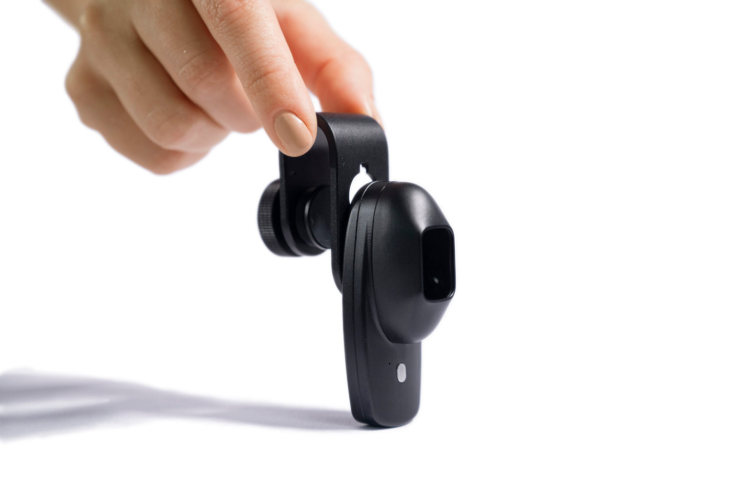
Since users do not generally have easy access to a dermatoscope with immersion, we have developed the lens shown in the photo, which allows viteye users to install it on their phone and, in tandem with high-quality cameras in modern phones, enables the full potential of the model to be revealed.
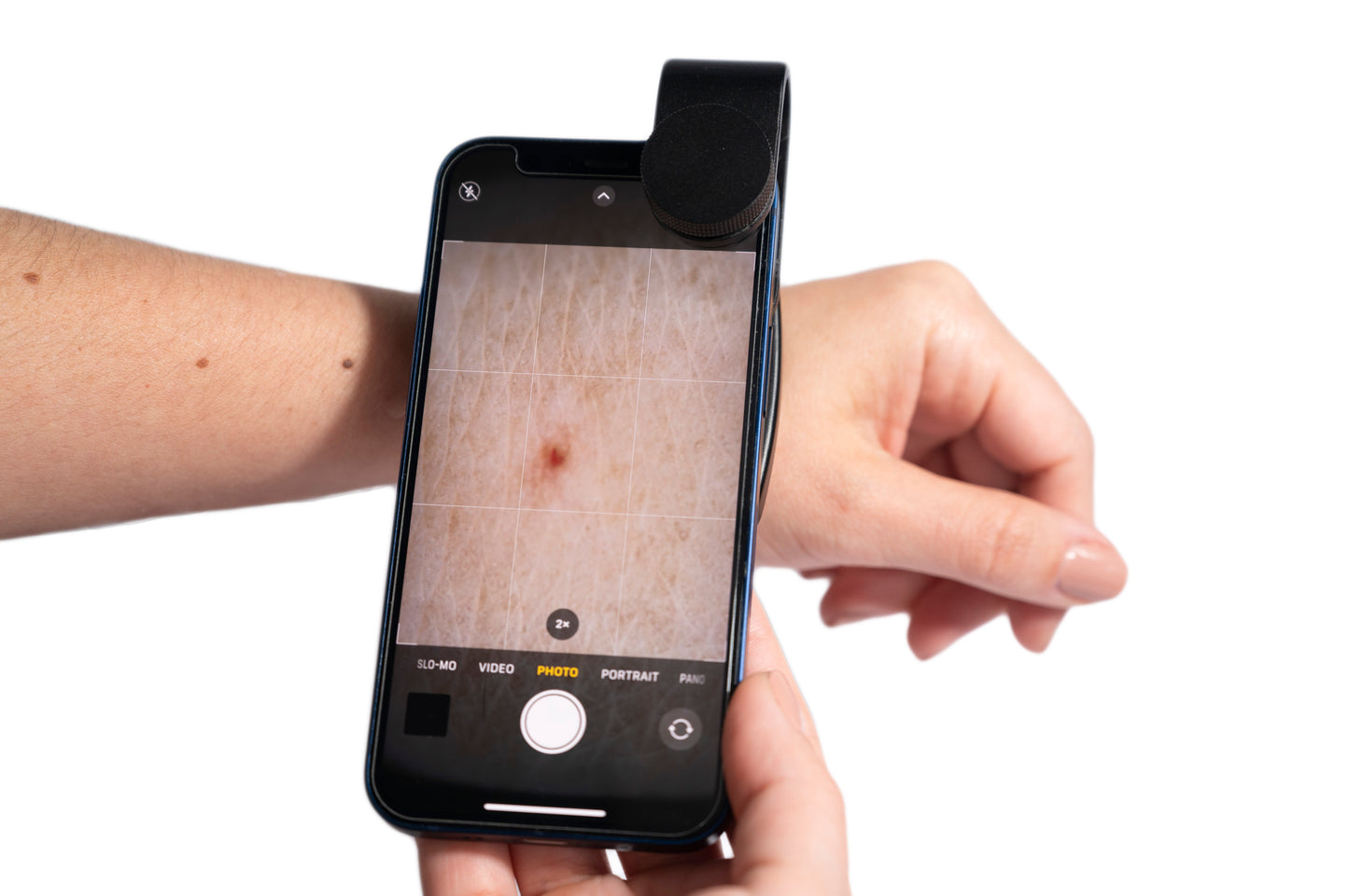
The lens attachment uses two cross-polarized LEDs, which allows the obtained image to be almost identical to the image obtained using liquid immersion. The polarized light eliminates skin glare and illuminates the upper layer of skin to obtain an image of a deeper structure of the neoplasm. It also allows for clearer, more precise, and detailed examination of the colors, shapes, and textures of skin lesions.
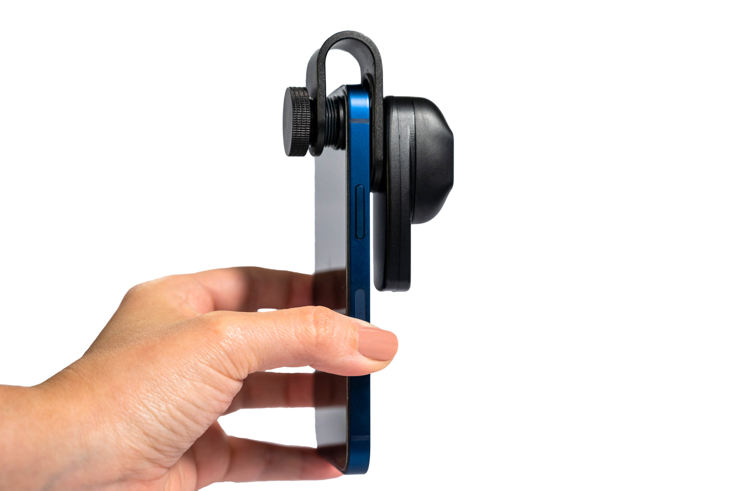
The device comes with a universal lens mount, which can be used to connect the dermatoscope to a smartphone or tablet for image capturing and display.
What’s in the Package
In the package, you will find the dermatoscope, universal lens mount, USB-C cable, carrying case, cleaning cloth, and user manual.
Operation
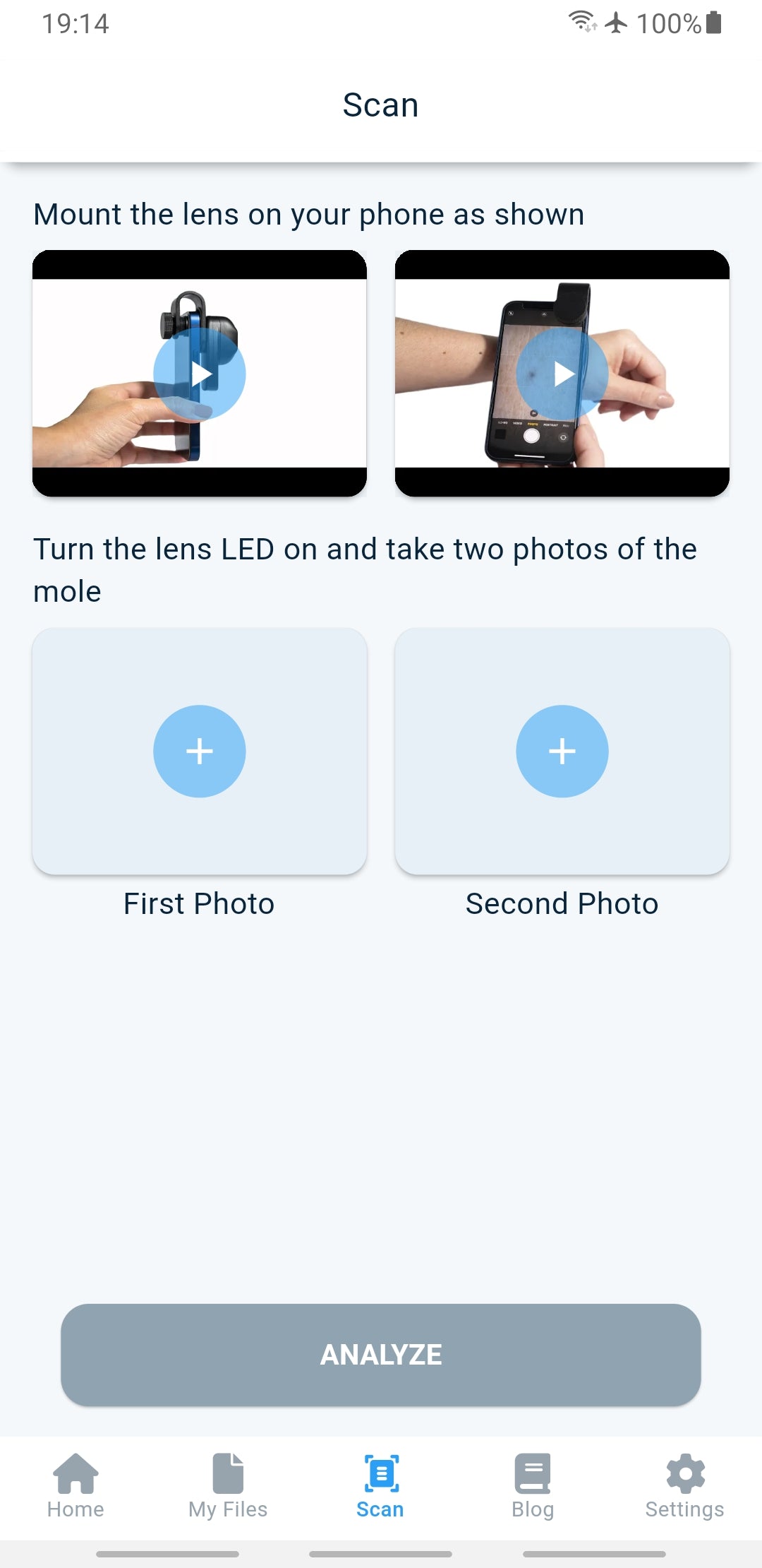
To operate the dermatoscope, you will need to attach it to a smartphone or tablet running viteye. First, align the lens mount with the center of the smartphone's camera. Next, align the dermatoscope with the mount and turn it 90 degrees to lock it in place. Turn on the polarized light by pressing the on/off switch.
Specs
Aluminum high quality shell.
All glass optical design. Low flare, broadband anti-reflection blue coating.
Cross-polarized light emitted by LED.
Battery capacity < 200 mAh. Working time: 1-2 hours.
Magnification: 4x.
Warranty
The device is maintenance-free and has a 2-year expected lifespan with proper use and adherence to warning and safety information, as well as maintenance instructions.
Disposal Instructions
When disposing of the device, it should not be treated as regular household waste. Dispose of the device at a facility that accepts electronics/electrical appliances.
Shipping from the US within the US.
Welcome to Viteye
Quick overview of the mounting procedure. Don't forget to take the phone case off!
Open Viteye, start a scan and take a photo with the dermatoscope. Easy as that!
Collapsible content
Return and Refund Policy
We have a 14-day return policy, which means you have 14 days after receiving your item to request a return.
To be eligible for a return, your item must be in the same condition that you received it, unworn or unused, with tags, and in its original packaging. You’ll also need the receipt or proof of purchase.
To start a return, you can contact us at support@viteye.ai
If your return is accepted, we’ll send you instructions on how and where to send your package. Items sent back to us without first requesting a return will not be accepted.
You can always contact us for any return question at support@viteye.ai.
Damages and issues
Please inspect your order upon reception and contact us immediately if the item is defective, damaged or if you receive the wrong item, so that we can evaluate the issue and make it right.
European Union 14 day cooling off period
Notwithstanding the above, if the merchandise is being shipped into the European Union, you have the right to cancel or return your order within 14 days, for any reason and without a justification. As above, your item must be in the same condition that you received it, unworn or unused, with tags, and in its original packaging. You’ll also need the receipt or proof of purchase.
Refunds
We will notify you once we’ve received and inspected your return, and let you know if the refund was approved or not. If approved, you’ll be automatically refunded on your original payment method within 10 business days. Please remember it can take some time for your bank or credit card company to process and post the refund too.
If more than 15 business days have passed since we’ve approved your return, please contact us at support@viteye.ai.
Terms and conditions
Terms of Use
Terms of use agreement
This Terms of Use Agreement (this “Agreement”) is entered into by and between the "Viteye" development team (the “Author”) and “you,” the user of this application, also known as the “Viteye'' (the “Application”). Access to, use of the Application is provided subject to the terms and conditions set forth herein. By accessing, using the Application, you hereby agree to these terms and conditions.
THIS AGREEMENT CONTAINS WARRANTY DISCLAIMERS AND OTHER PROVISIONS THAT LIMIT THE AUTHOR’S LIABILITY TO YOU. PLEASE READ THESE TERMS AND CONDITIONS CAREFULLY AND IN THEIR ENTIRETY, AS USING, ACCESSING THE APPLICATION CONSTITUTES ACCEPTANCE OF THESE TERMS AND CONDITIONS. IF YOU DO NOT AGREE TO BE BOUND TO EACH AND EVERY TERM AND CONDITION SET FORTH HEREIN, PLEASE EXIT THE APPLICATION AND DO NOT USE AND/OR ACCESS.
BY REGISTERING IN THE APPLICATION, YOU ACKNOWLEDGE AND AGREE THAT YOU HAVE READ AND UNDERSTAND THESE TERMS AND CONDITIONS, THAT THE PROVISIONS, DISCLOSURES AND DISCLAIMERS SET FORTH HEREIN ARE FAIR AND REASONABLE, AND THAT YOUR AGREEMENT TO FOLLOW AND BE BOUND BY THESE TERMS AND CONDITIONS IS VOLUNTARY AND IS NOT THE RESULT OF FRAUD, DURESS OR UNDUE INFLUENCE EXERCISED UPON YOU BY ANY PERSON OR ENTITY.
MEDICAL ADVICE DISCLAIMER
The Author provides the Application and the services, information, content and/or data (collectively, “Information”) contained therein for informational purposes only. The Author does not provide any medical advice in the application, and the Information should not be so construed or used. Using, accessing and/or browsing the Application and/or providing personal or medical information to the Author does not create a physician-patient relationship between you and the Author. Nothing contained in the Application is intended to create a physician-patient relationship, to replace the services of a licensed, trained physician or health professional or to be a substitute for medical advice of a physician or trained health professional licensed in your state. You should not rely on anything contained in the Application, and you should consult a physician licensed in your state in all matters relating to your health. You hereby agree that you shall not make any health or medical related decision based in whole or in part on anything contained in the Application.
INFORMATION DISCLAIMER
The information provided by our app and its features, including the lens, is for informational purposes only. Our app and lens are not a substitute for professional medical advice, diagnosis, or treatment. A proper diagnosis can only be made by a licensed medical professional. We recommend that users seek the advice of a physician or other qualified healthcare provider with any questions regarding personal health or medical conditions. The use of our app and lens does not create a physician-patient relationship between us and the user. Users are responsible for the proper handling and use of the lens. We are not responsible for any damage or injury caused by the misuse of the lens. Reliance on any information provided by our app or its features is solely at your own risk.
The opinions expressed in the Application are not necessarily the opinions of the Author and do not necessarily reflect the opinion of his employer(s). The Application is created by the Author in the Author’s individual capacity, is the Author’s personal application.
Any opinions of the Author on the Application are or have been rendered based on specific facts, under certain conditions, and subject to certain assumptions, and may not and should not be used or relied upon for any other purpose, including, but not limited to, for use in or in connection with any legal proceeding.
The Information may be changed without notice and is not guaranteed to be complete, correct, timely, current or up-to-date. Similar to any informational materials, the Information may become out-of-date. The Author undertakes no obligation to update any Information in the Application; provided, however, that the Author may update the Information at any time without notice in the Author’s sole and absolute discretion. The Author reserves the right to make alterations or deletions to the Information at any time without notice.
MESSAGING GUIDELINES
The Application is open to the public. Therefore, consider your messages in the chat carefully. By uploading or otherwise making available any information to the Author in the form of user generated messages or otherwise, you grant the Author the unlimited, perpetual right to distribute, display, publish, reproduce, reuse and copy the information contained therein.
You are responsible for the content you send in a message. You may not send content that is obscene, defamatory, threatening, fraudulent, invasive of another person’s privacy rights, or is otherwise unlawful. You may not post any content that contains any computer viruses or any other code designed to disrupt, damage, or limit the functioning of any computer software or hardware.
By submitting or posting content on the Application, you grant the Author and any company substantially under the control of the Author, the right to remove any content or comment that, in Author’s sole judgment, does not comply with the terms and conditions of this Agreement or is otherwise objectionable. You also grant the Author and any company substantially under the control of Author the right to modify, adapt, and edit any content.
THIRD PARTY LINKS AND ADVERTISEMENTS DISCLAIMER
The Application may, from time to time, contain links to third party web sites. These links are provided solely as a convenience to you and not as a guarantee, warranty, or recommendation by the Author of the services, information, content and/or data on such third party web sites or as an indication of any affiliation, sponsorship or endorsement of such third party web sites. The Author is not responsible for the content of linked third party web sites and does not make any representations or warranties regarding the privacy practices of, or the content or accuracy of materials on, such third party websites. If you decide to access linked third-party web sites, you do so at your own risk. Your use of third-party websites is subject to the terms of use for such sites.
THE INCLUSION OF THIRD PARTY ADVERTISEMENTS DOES NOT CONSTITUTE AN ENDORSEMENT, GUARANTEE, WARRANTY, OR RECOMMENDATION OF, AND THE AUTHOR MAKES NO REPRESENTATIONS AND/OR WARRANTIES ABOUT, ANY PRODUCT OR SERVICE CONTAINED THEREIN.
DISCLAIMER OF ALL WARRANTIES
The Information made available in the Application is provided on an “AS IS” and “AS AVAILABLE” basis without warranties of any kind, either express or implied, including, without limitation, warranties of title, noninfringement, and implied warranties of merchantability or fitness for a particular purpose. Without limiting the generality of the foregoing, the Author makes no warranty, representation or warranty as to the content, sequence, accuracy, timeliness or completeness of the Information, that the Information may be relied upon for any reason or that the Information will be uninterrupted or error free or that any defects can or will be corrected.
Without limiting the generality of the foregoing, the Author makes no representations or warranties with respect to any Information offered or provided within or through the Site regarding treatment of medical conditions, action, or application of medication.
Under no circumstances, as a result of your use of the Application, will the Author be liable to you or to any other person for any direct, indirect, special, incidental, exemplary, consequential or other damages under any legal theory, including, without limitation, tort, contract, strict liability or otherwise, even if advised of the possibility of such damages. Without limiting the generality of the foregoing, the Author shall have absolutely no liability in connection with the Application for:
- damages as a result of lost profits, loss of good will, work stoppage, failure of performance, delays in operation or transmission, nondelivery of information, deletions of files, mistakes, defects, errors, interruptions or computer failure or malfunction;
- any loss or injury caused, in whole or in part, by the Author’s actions, omissions, or negligence, or for contingencies beyond the Author’s control, in procuring, compiling, or delivering the Information;
- any errors, omissions, or inaccuracies in the Information regardless of how caused, or delays or interruptions in delivery of the Information; or
- any decision made or action taken or not taken in reliance upon the Information.
INDEMNIFICATION
You agree to indemnify and hold the Author harmless from any claim or demand, including attorneys’ fees, made by any third party as a result of (1) any content posted or made available by you in this Application, (2) any violation of law that occurs by you through the Application, and/or (3) anything you do using the Application and/or the Information contained therein.
INVALIDITY
If any provision of this Agreement is held to be invalid or unenforceable in whole or in part in any jurisdiction, then that provision shall be deemed ineffective in such jurisdiction but shall have no effect on the enforceability of the remaining provisions.
CONSENT TO JURISDICTION AND LIMITATION ON CLAIMS
You further agree that any claims or causes of action arising out of or related to this Agreement and the Application, along with the Information contained therein, shall be filed within one (1) year after such claim or cause of action arose, or such claim or cause of action shall be forever barred.
ENTIRE AGREEMENT
You hereby acknowledge that this Agreement represents the entire understanding between you and the Author concerning your use of the Application and the Information contained therein.
MODIFICATION
The Author may, in the Author’s sole and absolute discretion, modify the terms and conditions of this Agreement in whole or in part at any time for any reason without any notice to you, whether prior or otherwise. Such modified terms and conditions shall supersede these terms and conditions and shall become binding when published online in the Application.
WAIVER
The Author’s failure to exercise or enforce any right or provision of this Agreement shall not be deemed to be a waiver of such right or provision.
THE APPLICATION AND THE INFORMATION CONTAINED THEREIN IS MADE AVAILABLE BY THE AUTHOR FOR EDUCATIONAL PURPOSES ONLY AND IS NOT INTENDED TO PROVIDE MEDICAL ADVICE. BY ACCESSING THE APPLICATION, YOU UNDERSTAND AND ACKNOWLEDGE THAT THERE IS NO PHYSICIAN-PATIENT RELATIONSHIP BETWEEN YOU AND THE AUTHOR. YOU FURTHER ACKNOWLEDGE YOUR UNDERSTANDING THAT THE SITE SHOULD NOT BE USED AS A SUBSTITUTE FOR COMPETENT MEDICAL ADVICE FROM A LICENSED PHYSICIAN IN YOUR STATE.
How Viteye Works
About viteye
by Yevgeny Neretin
Medical curator, viteye
Candidate of Medical Sciences
Associate Professor (according to the Higher Attestation Commission)
Senior Oncology Specialist Doctor
by Yaroslav Sychov
Founder, viteye
In modern clinical oncology, the timely and early diagnosis of melanoma remains a significant and complex problem (Dinnes J, Deeks JJ, 2018; Holmes GA, Vassantachart JM et al., 2018; Dinnes J, Deeks JJ, Chuchu N, 2018).
Melanoma differs from other skin neoplasms due to its highly malignant course and, often, late diagnosis. In most cases, tumor treatment begins when it has already spread beyond the dermis (Satheesha T. Y., Satyanarayana D., et al., 2017). Consequently, melanoma has the highest mortality rates among other skin neoplasms.
The incidence of melanoma is increasing in many countries. While economically developed countries such as the USA and Germany have seen little to no change in melanoma mortality or even a slight decrease due to early diagnosis and timely treatment, most other countries observe an increase in mortality from melanoma (Apalla Z., et al., 2017; Ma J, Guo W, Li C, 2017; Kaprin A.D. et al., 2013).
In the United States, the incidence of melanoma has significantly increased over the past two decades. Between 2002 and 2009, the overall incidence of melanoma increased by about 4%. Notably, there was a more significant increase in the incidence of melanoma detected at the in situ stage (approximately 10%), while the number of cases of melanoma detected at the invasive stages even decreased (Linos E, Swetter SM, et al., 2009; Weinstock MA, Lott JP, et al., 2017; Clarke CA, McKinley Metal., 2017). This trend suggests the effectiveness of programs aimed at educating the population about early signs and the importance of preventing tumor development through measures like solar radiation protection (Grossman DC, Curry SJ, 2018).
In Norway, the incidence of melanoma has increased sevenfold over the past 50 years, making it one of the highest in the world (Robsahm TE, Bergva G, 2013). The authors believe that this rapid increase may be partly attributed to the fact that residents of this northern country have started traveling more and visiting southern countries.
Over a 25-year period from 1989 to 2013, the incidence of skin melanoma in most countries in the Mediterranean basin increased by 5.0% in men and 4.6% in women (Barbaric J, Laversanne M, et al., 2017). The rise in incidence is associated with the thinning of the ozone layer and an increase in the intensity of ultraviolet radiation. Several studies indicate an increasing incidence of melanoma in white people residing in countries geographically closer to the equator, where the proportion of ultraviolet radiation in sunlight is the highest and the number of sunny days per year is also high (Pinault L., et al., 2017; Liu-Smith F, et al., 2017).
To improve the accuracy of diagnosis, eliminate the influence of subjective factors, and reduce the time and cost of examining patients, special automated programs can be helpful. These programs can comprehensively assess the risk of developing melanoma in each specific case and analyze visual or dermatoscopic images of neoplasms. Although attempts to create such algorithms are being made, the available sample programs have not yet been widely used (Mar VJ, Scolyer RA et al., 2017).
Why did we take on this project?
We embarked on this project because the incidence of skin melanoma is increasing globally, and the knowledge of primary care specialists alone is often insufficient to make accurate decisions. Our goal in this project was to integrate modern technologies, patients, and qualified specialists to achieve early diagnosis of the disease.
Who is this product intended for?
This software product is intended for doctors, patients, and individuals seeking to enhance their expertise in the diagnosis of skin melanoma.
Use by doctors
Oncologists, dermatologists, and general practitioners who encounter pigmented neoplasms infrequently in their practice can utilize this software to support their decision-making process. For instance, a doctor can capture an image of a suspicious pigmented neoplasm and send it to a server for analysis. The software package will compare it with existing data in the database and provide the probability of a match in percentage. Ultimately, the doctor makes the final decision. In complex cases, doctors can initiate consultations on the platform and obtain opinions from other medical professionals.
Use by patients and healthy individuals
Patients and even healthy individuals can use the software to assess the "similarity" of their moles to melanoma. This can be particularly useful after a vacation when any growing neoplasm is detected. The patient may involve a doctor or a group of doctors to aid in the diagnosis.
What is not intended!
While the software package conducts accurate classification of images, it is important to note that the ultimate decision on further actions should ONLY BE MADE BY A DOCTOR! This includes the establishment of an early diagnosis of skin melanoma and the prescription of treatment. The software is primarily intended for educational purposes, drawing attention to the problem and encouraging potential patients to seek medical attention for suspicious neoplasms. However, it should NOT be relied upon as infallible. Although in some cases, the software may exceed the accuracy of a qualified oncologist, it is not flawless.
Limitations and Possible Errors of Image Classification
The program may misclassify your skin image due to the following reasons:
- Insufficient sharpness or blurry image with poor quality.
- Atypical presentation of the disease, which is extremely rare. For example, a site of malignancy on the edge of a nevus or non-pigmented melanoma. In such cases, even qualified dermatologists or oncologists can make mistakes, and the correct diagnosis can only be made after the removal of the neoplasm and subsequent histological examination.
- Presence of hair, artifacts, or other "interference" in the image.
Creation, Mechanism, Internal Structure, and Principle of Operation
Software Part:
The complex operates based on neural networks.
The essence of the system lies in the utilization of neural networks for diagnosing skin melanoma, where a specific pathology is diagnosed using a computer neural network model.
To achieve this goal, we had to address several specific tasks, including:
- Determining the optimal volume of training and testing samples.
- Identifying the optimal type of neural network.
- Finding the optimal architecture for the selected network type.
- Evaluating the effectiveness and significance of the created model.
- The program is based on a trained neural network, which provides the probability of melanoma in a given patient as a result.
Training a neural network in most standard cases is an automatic process, which requires specialist involvement only after its completion to evaluate the results. In our case, the training process took approximately 10-12 hours of machine time. The system's performance was evaluated through testing using a control sample at any stage of training.
Interface Development:
An interface was created to develop a user-friendly and efficient computer system that is accessible for use by doctors without specialized technical education.
Debugging and Testing:
This stage involved debugging the program and bringing the testing to a real system for practical healthcare applications.
Refinement:
This stage is characteristic of learning systems. After the initial training stage, the system can continue learning in real work conditions and incorporate new real data. The fundamental difference between creating neural network systems and traditional data processing methods lies in the fact that the system is never instantly ready or completely finished. It continues to accumulate experience during operation.
We took this project seriously.
In the initial stage, a training sample was formed, consisting of 6,144 clinical cases of patients who underwent examination and surgical treatment, and whose diagnoses were histologically verified. The collected material amounted to a total of 16 gigabytes of images. This data was gathered as part of a dissertation research for the degree of Doctor of Medical Sciences conducted between 2012 and 2023.
In the second stage, the neural network is trained using the provided training data. To determine the optimal type of network, training is carried out on several standard models.
These include, at a minimum, a linear network, a multilayer perceptron, and a network with a radial basis function. The selection program automatically chooses the best models from the set of created models. The optimal network architecture is determined through empirical experimentation.
The best model is selected based on the ratio of standard deviations, which is the ratio of the standard deviation of the forecast error to the standard deviation of the input data. The model is considered successful if the standard deviation ratio approaches zero. The fraction of the model variance explained is calculated as one minus the ratio of standard deviations.
The criterion for successful learning is the progressive reduction in error on the training set. This error is computed as the total squared deviation between the values at the outputs of the neural network in the training sample and the real values obtained at the outputs of the neural network.
The learning process is stopped if the error on the control set increases while it continues to decrease on the training set. This indicates overfitting of the network, where the network has learned the training sample too precisely, resulting in reduced diagnostic quality when new data is fed into the network.
In the third stage, the created model is tested by comparing the obtained values with a set of known data that was not used during training and testing. This evaluation assesses the quality of the diagnosis and the effectiveness of the resulting model.
Hardware
To enhance the accuracy of diagnosing skin melanoma, we have developed a hardware solution. Our goal during the development process was to achieve the highest possible accuracy while keeping the patient's investment relatively minimal.
Currently, visual examination is the primary method for diagnosing melanoma. However, evidence suggests that some melanomas may be missed if visual examination is used without additional diagnostic methods, such as digital dermatoscopy (Dinnes et al., 2018; Dinnes et al., 2018).
With appropriate training, doctors from various specialties can often provide a preliminary diagnosis of "skin melanoma" during the initial patient examination (Fink & Haenssle, 2017).
Several mnemonic rules exist for diagnosing melanoma and identifying high-risk precancerous neoplasms based on visual examination, patient questioning, complaint collection, and medical history. Commonly used rules include the ABCDE rule, Figaro rule, Glasgow rule, DOCTOR rule, and others. These rules aid first-contact doctors in quickly and accurately diagnosing skin melanoma and help them memorize the symptoms necessary for diagnosing a malignant tumor. However, it has been noted that these rules do not provide sufficient diagnostic accuracy for early-stage tumors (melanoma in situ).
One of the well-known diagnostic symptom complexes is the ABCD rule proposed by R. Friedman in 1985.
This rule assesses pigmented skin neoplasms based on four parameters:
A (asymmetry) - asymmetry of the pigment spot;
B (border) - irregularity of the border;
C (color) - uneven coloration;
D (diameter) - diameter exceeding 6 mm.
Later, the rule was expanded to include criterion E (evolution), which evaluates dynamic monitoring of individuals at risk and allows for the assessment of changes in the color, shape, and size of the pigmented neoplasm.
The diagnostic accuracy is significantly increased when the ABCD rule is used in conjunction with digital dermatoscopy (Demidov & Sokolov, 2007). The doctor's experience also plays a significant role (Naldi & Falgheri, 2018), and dermoscopy training helps clinicians improve their ability to diagnose melanoma (Secker et al., 2017; Grange & Woronoff, 2014; Xavier & Drummond-Lage, 2016).
But how can we ensure that patients have access to early diagnosis at the level of a qualified specialist from the comfort of their homes? We have found a solution. We developed a lens with a polarization mode that, when used in conjunction with a modern smartphone's high-quality camera, allows for the visualization of subcutaneous structures and identification of minimal dermatoscopic elements. This lens, combined with a glass magnifying lens featuring a special coating and cross-polarization LEDs, enables the correct diagnosis by classifying the observed object.
Indicators of Diagnostic Performance of the Developed Model
We have achieved network performance indicators that are in line with the findings of other researchers in the field.
Expert systems based on neural networks have emerged, enabling accurate diagnosis of melanoma at the level of qualified dermatologists and real-world testing (Brinker et al., 2019; Tschandl et al., 2019). In many cases, these systems have demonstrated higher accuracy compared to doctors from other specialties, such as surgeons (Mar et al., 2018; Oakden-Rayner, 2018).
Of particular interest is the study by Tschandl et al., which showed that while highly qualified dermatologists and the program achieved similar levels of diagnostic sensitivity (approximately 92%), the computer-based system exhibited higher diagnostic specificity (61.1%) compared to doctors (57.7%) (Tschandl et al., 2019).
We have calculated the sensitivity and specificity of our program.
Sensitivity refers to the proportion of truly diseased individuals in the tested population who are correctly identified as sick by the diagnostic test. It is a measure of the likelihood that the test will correctly detect instances of the disease. In the context of melanoma, sensitivity indicates the program's ability to avoid missing cases.
Specificity, on the other hand, refers to the proportion of non-diseased individuals who test negative for the disease among the entire population without the condition. It is a measure of the test's ability to correctly identify individuals without the disease. In simple terms, if there is no illness present, the program should correctly identify it as such.
To provide a better understanding of the value of our system, here are some important facts:
Melanoma is relatively rare in routine medical practice, including among oncologists. The clinical presentation of early forms of melanoma is often uncertain, making interpretation of the signs challenging due to limited knowledge. As a result, first-contact doctors sometimes make errors when faced with cases of pigmented skin neoplasms (Dimov, 2008).
These errors can be categorized into two main groups: type I errors, associated with underdiagnosis, and type II errors, leading to overdiagnosis of neoplasms (Dimov, 2008).
Type I errors pose greater risks to the patient's life, as they directly impact treatment outcomes and prognosis.
However, type II errors in melanoma diagnosis are also unacceptable, as they can lead to unnecessary surgical interventions, significant cosmetic defects, and reduced quality of life (Zaridze & Maksimovich, 2017; Sokolova & Malishevskaya, 2018).
Additionally, situational or incidental errors may occur due to atypical disease progression or comorbidities, where certain diseases significantly distort the presentation of others (Ponkina, 2012; Ludupova, 2016).
Privacy Policy
Privacy Policy for viteye
Last Updated: 25 May, 2024
Thank you for using viteye, an app designed to provide insights on photographs using a trained neural network. Viteye aims to assist in the early detection of melanoma through educational information. Please note that while viteye's neural network has been trained on thousands of clinical photographs, it is not a substitute for medical advice. Consult a qualified healthcare provider for medical advice if needed.
Viteye is committed to protecting your privacy and providing a secure and reliable service. We respect your personal information and are transparent about the data we collect and how we use it.
Data Collection:
When you use viteye, we collect certain personal information from you, such as your name, age, gender, height, weight, skin type, hair color, eye color, presence of freckles, type of labor,tanning bed experience, smoking status, involvement in hazardous production and the level of solar activity in your place of residence.
This information is used to determine the risk of melanoma and to provide more appropriate information.
We also collect photos of moles or other skin lesions that you upload to viteye for analysis. These photos are securely stored in our database and are used solely for the purpose of determining the risk of melanoma. The photos are not linked to your personal identity, and we do not use them for any other purposes.
The collection of this information is entirely voluntary, and providing accurate details is not mandatory. Users can also choose to provide information anonymously.
We do not collect any sensor data, and only the photographs taken with the camera will leave the user's device to be uploaded to our server. The photographs will be visible to the doctors during checkups.
In addition, we may collect information about your device, such as your device type and operating system. This information is used to improve viteye and to ensure that it is compatible with your device.
Our app is not intended for children, and we do not collect any information from children.
Data Sharing:
None of the user information is shared with any third parties except for the photographs uploaded to a third-party server for neural network analysis. The photographs are stored encrypted and anonymously, without any link to the user. Uploaded photographs may be used to train the neural network for future improvements.
We may share your personal information and photos with medical professionals who use viteye to provide appropriate information. We may also share your personal information with other third parties if required by law or to protect our legal rights.
However, we will not share your personal information or photos with any third party for marketing or advertising purposes.
Data Security:
We take data security seriously and have implemented appropriate technical and organizational measures to protect your personal information from unauthorized access, use, or disclosure. We use industry-standard encryption to protect your data in transit and at rest.
We also limit access to your personal information to only those employees and contractors who need to know it for the purpose of providing our services. All of our employees and contractors are bound by strict confidentiality agreements and are required to follow our data security policies.
Data Accuracy:
We rely on the accuracy of the information you provide to us in order to provide accurate information. We encourage you to provide us with complete and accurate information, but we understand that mistakes can happen. If you need to update or correct any of your personal information, please contact us at support@viteye.ai .
Data Retention:
Users have the option to delete their data completely by deleting their account from the app. Upon account deletion, all private information, along with the associated photographs, will be removed from our servers, unless we are required by law to retain them.
We will retain your personal information and photos for as long as necessary to provide our services to you, unless a longer retention period is required by law.
Changes to our Privacy Policy:
We reserve the right to update or change our Privacy Policy at any time. Any changes will be posted on this page, and we will notify you if any material changes are made.
Contact Us:
If you have any questions or concerns about our Privacy Policy or the way we collect and use your personal information, please contact us at support@viteye.ai .
This version of the privacy policy was created on May 25, 2024 . Updated versions will be sent via email to users registered in the app.
Contact Us
Feel free to contact us if you have any questions regarding the lens or the Viteye application.
Email: support@viteye.ai
Phone: + 1 918 309 2022
Viteye OÜ
Estonian limited company number 16766170.
Veskiposti 2-1002
Tallinn 10138, Harju maakond
Estonia


